Sherrilyn Phelps, P.Ag. and Dr. Barb Zeisman, A.Ag.
Pathogens Involved
Pseudomonas syringae pv. pisi is the main pathogen involved. Bean bacterial brown spot (Pseudomonas syringae pv. syringae) can also attack peas, causing similar symptoms, but is less common in Western Canada.
Occurrence
Bacterial blight occurs worldwide. Samples of peas have been identified with moderate to severe symptoms of bacterial blight by the North Dakota State University Plant Pathology Department following periods of wet conditions in 2015, but it is not considered an economically important disease of peas. In Western Canada it is considered to have minimal yield impact on peas.
In 2017, bacterial blight symptoms were identified in Alberta pea fields. DNA-based testing (PCR) was used to confirm the diagnosis and identified Pseudomona syringae pv pisi as the causal organism. Bacterial blight was only observed early in the season (mid-June) and disease progression to the upper canopy and pod lesions were rarely observed.
Since 2017, there have been fields identified with bacterial blight symptoms across the Prairies provinces. The prevalence of this disease is not constant and is largely driven by environmental conditions. Through most surveys, disease diagnosis is made based on visible symptoms and the specific pathogen involved has not always been determined. For accurate diagnosis, DNA-based testing is required.
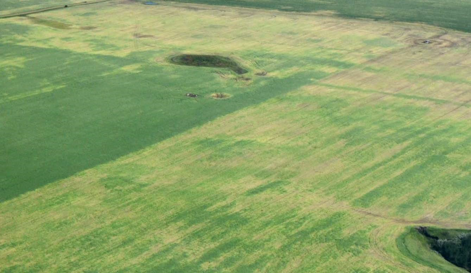
If the pathogen is introduced via infected seed symptoms are often identified in localized areas in the field, whereas the disease may be more widespread in a field if the primary inoculum originates from infected residue within the field.
Symptoms

Sources: Dr. Syama Chatterton, Agriculture and Agri-Food Canada Lethbridge, R.A.A. Morrall, University of Saskatchewan
Source: Dr. Syama Chatterton, Agriculture and Agri-Food Canada Lethbridge, AB
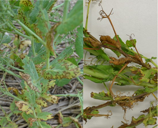
Symptoms of seed-borne infections may appear first as small water-soaked oval spots on the lower parts of the plant such as stems, leaves, and stipules. Bacterial ooze may be visible within the lesions under high humidity.
Figures 1 and 2 show close ups of individual lesions as well as the whole plant symptoms in the field. The water-soaked lesions on stiplules and leaves may enlarge and coalesce, but are often limited by the veins creating an angular appearance. Older lesions have dark margins with brown and papery (translucent) centre.
Lesions on the stems begin as water-soaked areas, which later turn olive-green to dark brown. Stem lesions may coalesce, causing the stem to shrivel and die (Figure 2). Stem infection may spread upwards to the stipules and leaflets. Infection of sepals can result in blossom abortion.
Infected pods will also have water-soaked circular lesions at first, but lesions become sunken and turn darker as they mature (Figure 3). The suture area is often a site of infection that can result in seed infection. Infected seed may or may not show symptoms. Heavily infected seed may be discoloured, but light infection has no visible effect on seed. If symptoms appear they tend to be watery, dark spots.
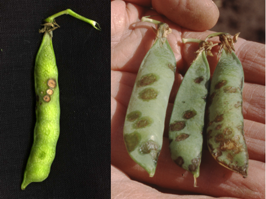
Sources: Dr. Banniza and R.A.A. Morrall, University of Saskatchewan
Symptoms of bacterial blight and Ascochyta (Mycosphaerella) blight are easily confused. Bacterial blight lesions are more angular and translucent (particularly when held up to light). Knowing which disease is present is important as fungicide applications will not control bacterial blight.
Bacterial blight may be confused with brown spot which is caused by P. syringae pv. syringae. The symptoms are difficult to distinguish in field situations. P. syringae pv. syringae is not common in Western Canada.
Disease Cycle
The disease commonly becomes established within a field by sowing infected seed. Once a field is infected, the pea residue can be a source of inoculum for future infections as well as infections in neighbouring fields.
Infected seed is covered by a dry, white film of bacteria, both externally and internally, that is invisible to the naked eye (Skoric 1927; Lawyer 1984). Seed can remain infected for up to three years. The plumule contacts the infected seedcoat during germination and infection of the seedling begins. When seed infection is the source of inoculum the lowest three stipules are the most common infection sites. Optimal temperature for growth are 26-28°C. The higher the soil moisture, the higher the rate of infection. Injury predisposes the plants to infection. Early infections are more damaging. Often infected seedlings do not survive.
During wet weather, secondary infection occurs as bacteria spread from infected to healthy plants by rain splash, wind-borne water droplets, and plant to plant contact. Infection may occur at any stage of plant growth and is most prevalent following frosts, hail damage or heavy rains that cause injury to otherwise healthy tissue. Therefore, it is more prevalent when frequent rains occur in combination with or following hail, frost, or heavy winds.
Environmental Factors
Cool, overcast weather with high humidity promotes disease development. Dry conditions and higher temperatures slow or stop the progression of bacterial blight.
Control
Plant disease-free seed – The bacterium is carried within infected seeds and current fungicide seed treatments have no impacted on controlling the spread of bacterial blight from infected seed.
Crop rotation – Minimum three-year rotation to avoid risk. A four-year rotation is recommended if disease is identified. Stubble can be a significant source of inoculum therefore it is important to allow residue to completely breakdown before replanting to peas.
Avoid damage from herbicides and physical damage (machinery).
Fungicide application – Typical chemical fungicides (seed treatments or foliar fungicides) are not effective as the pathogen is bacterial – not fungal. However, copper-based fungicides do have some antibacterial properties but are only registered for brown spot (Pseudomonas syringae pv. Syringae) control on peas. Little information is available on the effectiveness at protecting yield or reducing seed infection.
References
- Chatterton, S., Harding, M., Bowness, R., Vucurevich, C., Dubitz, V., Nielsen, J., Gossen, B., Banniza, S., Ziesman, B. and McLaren, D. 2018. Survey for diseases of pulse crops in Alberta, Saskatchewan and Manitoba in 2017. Western Committee on Plant Disease Pulse Disease Situation Report and Research Update. Pg 12-14
- Hadedorn, D.J. 1991. Handbook of Pea Diseases.
- Harding, M., Chatterton, S., Bowness, R., Vucurevuch, C., Dubitz, T., Daniels, G. and Burke, D. 2018. Survey for disease of pulse crops in Alberta, Saskatchewan and Manitoba in 2018. Western Committee on Plant Disease Pulse Disease Situation Report and Research Update. Pg 21-22.
- Laflamme, P. 1986. Disease of Field Pea (Pisium sativum L.) in the Peace River Region of Alberta. MSc thesis.
- Ludwig, C. A. 1926. Pseudomonas (Phytomonas) pisi Sackett, the cause of a pod spot of garden pea. Phytopathology. 16:177-183.
- McLaren, D.L., Kim, Y.M., Conner, R.L., Derksen, H., Chang, K.F., Hwang, S.F., Henderson, T.L., Penner, W.C., Thompson, M.J., Stoesz, D.B. and Kerley, T.J. 2017. 2017 disease surveys of dry bean, field pea and soybean in Manitoba. Western Committee on Plant Disease Pulse Disease Situation Report and Research Update. Pg 5-6
- McLaren, D.L., Kim, Y.M., Conner, R.L., Derksen, H., Chang, K.F., Hang, S.F., Henderson, T.L., Penner, W.C., Thompson, M.J., Stoesz, D.B., Kerlerl, T.J., Stevenson, L., Tkachuk, C., and Brown-Livingston, K. 2018. 2018 root disease surveys of dry bean, field pea, and soybean and phytophthora root rot in Manitoba. Western Committee on Plant Disease Pulse Disease Situation Report and Research Update. Pg 4-5.
- North Dakota State University. Crop and Pest Report. 2015. Bacterial blight of Peas in North Dakota and Minnesota:
- Skoric V. 1927. Bacterial blight of pea: overwintering, dissemination and pathological histology. PhytopathoIogy. 1 7:6 1 1-627
